- Overview
- Log In For Videos
- Give Feedback
- Seizure Classification
- Unknown Onset Seizure
- Neonatal Seizure
- Epilepsy Classification
- Generalized Epilepsy
- Focal Epilepsy
- Generalized and Focal Epilepsy
- Unknown Epilepsy
- Epilepsy Syndromes
- Epilepsy Etiologies
- Metabolic Etiologies
- Immune Etiologies
- Infectious Etiologies
- Unknown Etiologies
- Encephalopathy
- Epilepsy imitators
DEVELOPMENTAL AND/OR EPILEPTIC ENCEPHALOPATHY WITH SPIKE-WAVE ACTIVATION IN SLEEP (DEE-SWAS, EE-SWAS)
Background
The background may be normal or there may be focal, multifocal or diffuse slowing, depending on the etiology. Normal sleep architecture is absent or difficult to distinguish due to epileptiform abnormality in sleep.
Interictal
Slow (1.5–2Hz) spike-wave discharges are markedly activated in non-REM (stage I-III) sleep. They may be seen diffusely, focally (typically frontally) or multifocally.
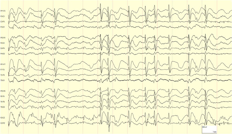
Example of continuous spike-wave in sleep.
CAUTION An overnight sleep EEG may be required, as spike-wave activation in sleep may not be seen during brief periods of sleep.
Activation
The EEG is activated in non-REM sleep.
Ictal
The ictal EEG pattern correlates with the seizure type seen.
Feedback
|
Home
|
Contact Us
|
Privacy
|
Terms & Conditions of Use
|
Log In For Videos

This website is owned by the International League Against Epilepsy. Text on this website, last updated June 30, 2024,
is available under a Creative Commons Attribution-ShareAlike 4.0 International License,
EXCEPTING all videos and images, which remain copyrighted by the International League Against Epilepsy.