- Overview
- Log In For Videos
- Give Feedback
- Seizure Classification
- Unknown Onset Seizure
- Neonatal Seizure
- Epilepsy Classification
- Generalized Epilepsy
- Focal Epilepsy
- Generalized and Focal Epilepsy
- Unknown Epilepsy
- Epilepsy Syndromes
- Epilepsy Etiologies
- Metabolic Etiologies
- Immune Etiologies
- Infectious Etiologies
- Unknown Etiologies
- Encephalopathy
- Epilepsy imitators
SELF-LIMITED EPILEPSY WITH AUTONOMIC SEIZURES (SeLEAS)
Background
The background EEG is normal.
CAUTION Focal slowing
consistently over one area is not seen  consider structural brain abnormality.
Interictal
A standard EEG can be normal in some patients. Multifocal high voltage spikes or sharp-waves are typically seen, these often are present in different focal areas on sequential EEGs. All focal brain regions may be affected but abnormality is often over the posterior (occipital) regions.
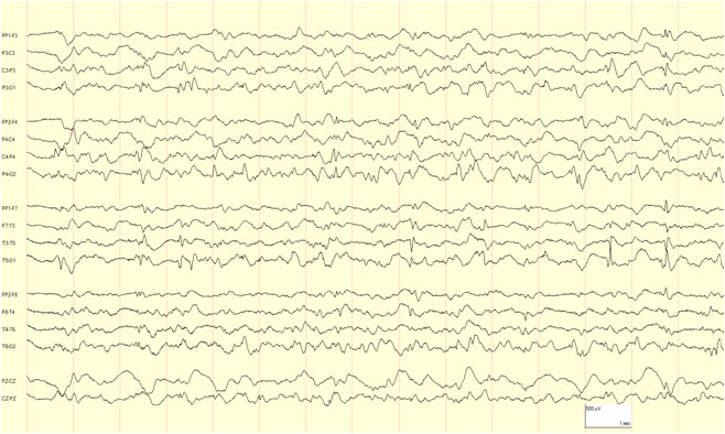
Example of multifocal spikes in self-limited epilepsy with autonomic seizures.
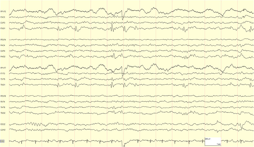
Example of occipital (left) spikes of high amplitude in self-limited epilepsy with autonomic seizures.
Activation
EEG abnormality is enhanced by sleep deprivation, in drowsiness and in sleep, when discharges often have a wider field and may be bilaterally synchronous. Eye closure (elimination of central vision and fixation off sensitivity) may activate posterior discharges in some patients.
Ictal
Ictal patterns are unilateral, often having posterior onset, with rhythmic slow (theta or delta) activity intermixed with small spikes and/or fast activity.
This website is owned by the International League Against Epilepsy. Text on this website, last updated June 30, 2024,
is available under a Creative Commons Attribution-ShareAlike 4.0 International License,
EXCEPTING all videos and images, which remain copyrighted by the International League Against Epilepsy.