- Overview
- Log In For Videos
- Give Feedback
- Seizure Classification
- Unknown Onset Seizure
- Neonatal Seizure
- Epilepsy Classification
- Generalized Epilepsy
- Focal Epilepsy
- Generalized and Focal Epilepsy
- Unknown Epilepsy
- Epilepsy Syndromes
- Epilepsy Etiologies
- Metabolic Etiologies
- Immune Etiologies
- Infectious Etiologies
- Unknown Etiologies
- Encephalopathy
- Epilepsy imitators
POLYMICROGYRIA
Imaging
Imaging for optimized detection of polymicrogyria:
MRI is the imaging of choice for assessing the detail and associated structural abnormalities. MRI should include thin slice volumetric T1-weighted images, axial and coronal T2-weighted and FLAIR images. Thin sliced imaging is necessary to distinguish the microgryia in the thickened cortex, which may otherwise resemble the thickened cortex and poorly formed gyral pattern seen in lissencephaly or the thickened cortex with grey-white matter blurring of focal cortical dysplasia.
Imaging characteristics of polymicrogyria:
- apparent cortical thickening due to multiple small gyri (over-folding, 6 to 10 mm thickness) rather than true thickening
- an irregular surface to the cortex due to multiple small gyri
- a stippled appearance to the grey-white matter interface due to multiple small gyri
- Sylvian fissue may show abnormal morphology, being extended and abnormally oriented posteriorly
- underlying white matter may show high signal on T2 weighted images
- occasionally, perhaps more so in patients with previous intrauterine infection, the abnormal cortex may show regions of calcification.
- abnormality of the basal ganglia suggests a tubulin gene pathogenic variant
- may be associated periventricular nodular heterotopia
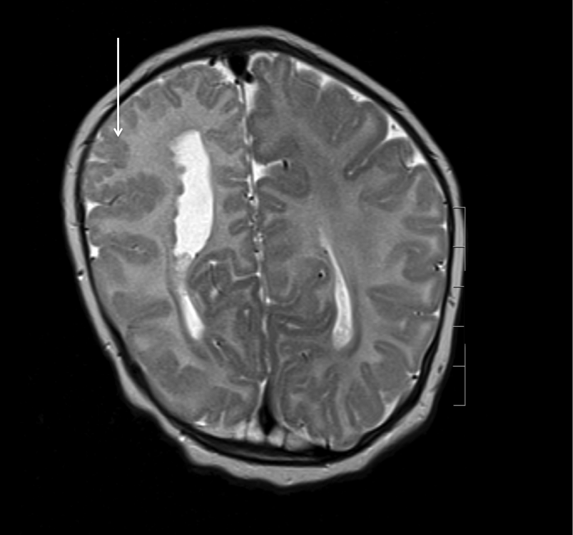
Imaging of polymicrogyria
The image below is from a patient with Aicardi syndrome and shows a localized area of polymicrogyria in the right frontal lobe (arrow)

Imaging of polymicrogyria
The image below shows unilateral (right) periventricular nodular heterotopia, with grey matter heterotopia lining the body of the right ventricle. There is polymicrogyria in the overlying cortex (arrow)
This website is owned by the International League Against Epilepsy. Text on this website, last updated June 30, 2024,
is available under a Creative Commons Attribution-ShareAlike 4.0 International License,
EXCEPTING all videos and images, which remain copyrighted by the International League Against Epilepsy.