- Overview
- Log In For Videos
- Give Feedback
- Seizure Classification
- Unknown Onset Seizure
- Neonatal Seizure
- Epilepsy Classification
- Generalized Epilepsy
- Focal Epilepsy
- Generalized and Focal Epilepsy
- Unknown Epilepsy
- Epilepsy Syndromes
- Epilepsy Etiologies
- Metabolic Etiologies
- Immune Etiologies
- Infectious Etiologies
- Unknown Etiologies
- Encephalopathy
- Epilepsy imitators
DNET
Imaging
Imaging for optimized detection of DNET:
MRI should include thin slice volumetric T1-weighted images, axial and coronal T2-weighted and FLAIR images.
Imaging characteristics of DNET:
- DNET's are typically cortical lesions that lack significant mass effect or peri-tumoral edema
- They are hypointense on T1-weighted images and hyperintense on T2-weighted images, typically with a 'bubbly appearance' due to their multicystic nature
- They may show rings of enhancement, seen on FLAIR ('bright rim sign')
- They may be associated with scalloping of the inner table of overlying skull bone, without actual skull bone erosion
- They may show calcification (seen in ~30%), this may be best seen on CT or T2* weighted MR imaging
DNET's can co-occur with focal cortical dysplasia (which is usually found adjacent to the DNET) and/or with hippocampal sclerosis.
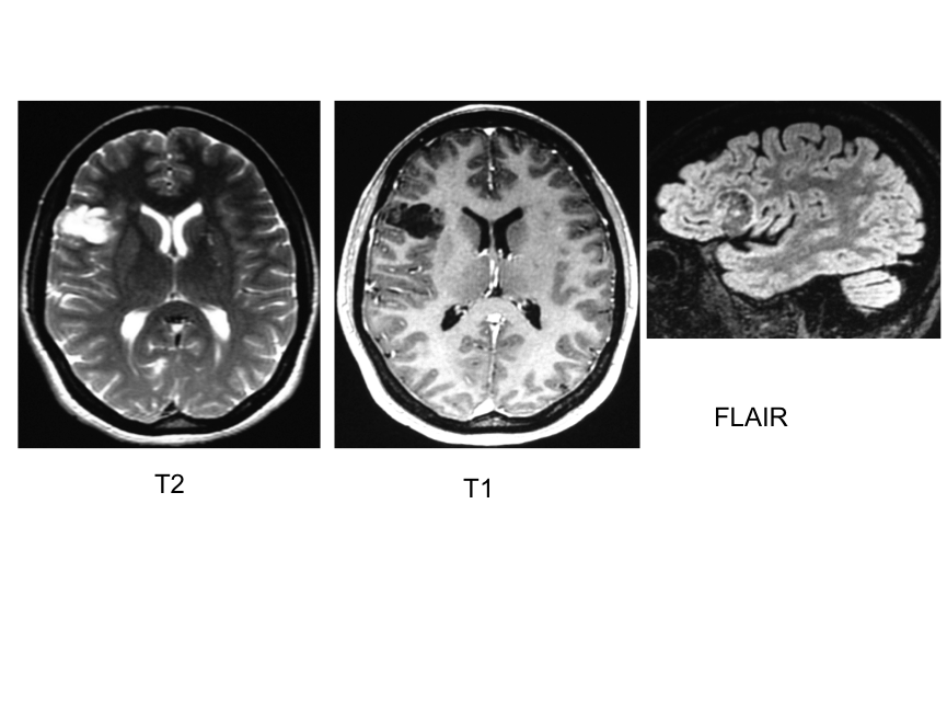
Imaging of a DNET
The three images below are a T2-weighted, T1-weighted and FLAIR image of a DNET in the same patient, demonstrating the multicystic appearance, with a 'bright rim sign' - a ring of enhancement, seen on the FLAIR image .
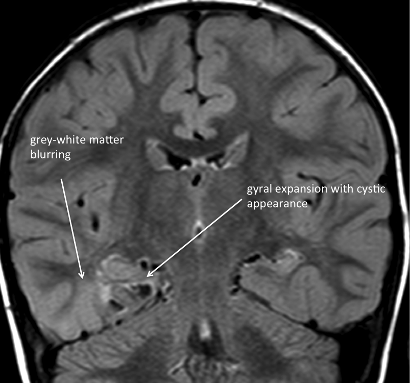
Imaging of a DNET with focal cortical dysplasia (FCD IIIb)
The image below is a coronal image showing a right basal temporal structural abnormality, which includes an area of gyral expansion, with cystic appearance, and neighboring abnormal signal in white matter, with blurring of the grey-white matter junction. This abnormality was confirmed following resection to be a DNET with adjacent focal cortical dysplasia (FCD IIIb).
This website is owned by the International League Against Epilepsy. Text on this website, last updated June 30, 2024,
is available under a Creative Commons Attribution-ShareAlike 4.0 International License,
EXCEPTING all videos and images, which remain copyrighted by the International League Against Epilepsy.