- Overview
- Log In For Videos
- Give Feedback
- Seizure Classification
- Unknown Onset Seizure
- Neonatal Seizure
- Epilepsy Classification
- Generalized Epilepsy
- Focal Epilepsy
- Generalized and Focal Epilepsy
- Unknown Epilepsy
- Epilepsy Syndromes
- Epilepsy Etiologies
- Metabolic Etiologies
- Immune Etiologies
- Infectious Etiologies
- Unknown Etiologies
- Encephalopathy
- Epilepsy imitators
STURGE-WEBER SYNDROME
Imaging
Imaging for optimized detection of Sturge-Weber syndrome:
While Sturge-Weber syndrome may be detected on skull Xray and CT, especially when there is established calcification and atrophy, MRI is the imaging of choice for assessing detail. MRI should include thin slice volumetric T1-weighted images with contrast, and axial and coronal T2-weighted and FLAIR images. In addition, orbital involvement can be assessed with orbital MRI.
Imaging characteristics of Sturge-Weber syndrome:
- On CT imaging, gyral subcortical calcification is seen, with a "tram-track" appearance, unilateral cerebral atrophy can also be appreciated.
- On MR imaging, T1-weighted imaging with contrast shows leptomeningeal enhancement, this may not be seen in older patients. Other findings include cerebral atrophy, white matter signal abnormality, and an enlarged unilateral choroid plexus.
- Angiography is not routinely performed, but is abnormal in most cases, showing absent superficial cortical veins, with dilated and tortuous deep veins.
CAUTION imaging findings may be absent in infants, as calcification and atrophy may be absent at this age.
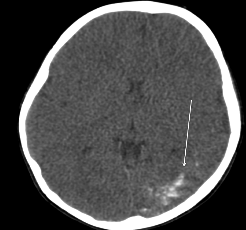
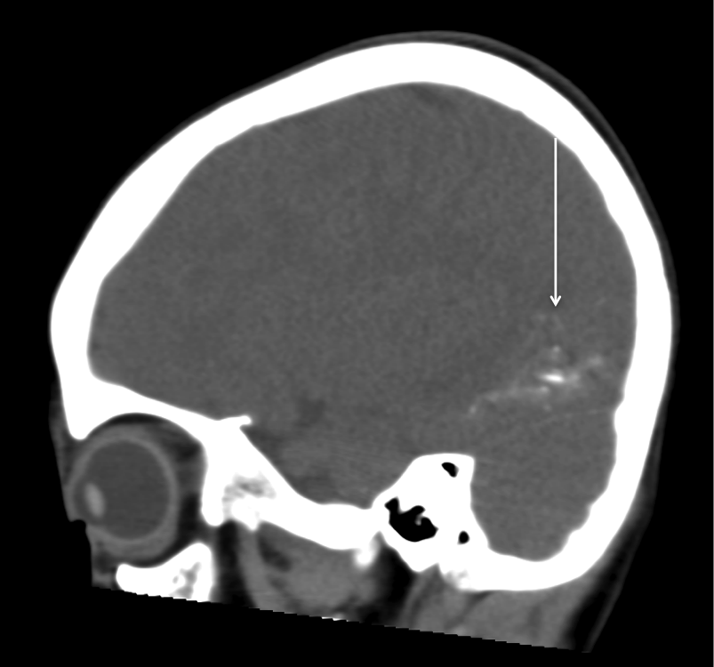
Imaging of Sturge-Weber syndrome
The two CT images below show subcortical calcification in the left occipital lobe (arrows).
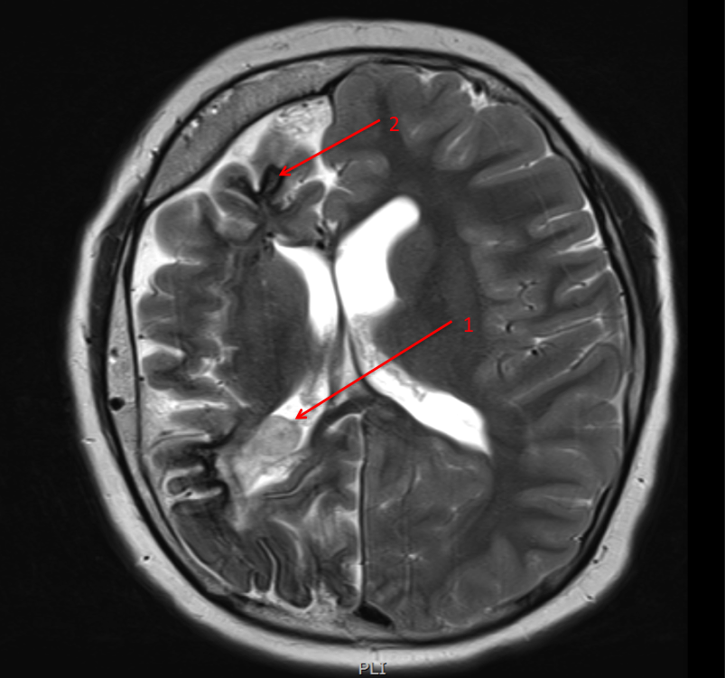
Imaging of Sturge-Weber syndrome
The image below is a T2 weighted image, showing atrophy of the right hemisphere, enlargement of the choroid plexus (arrow 1), a thickened overlying skull and subcortical calcification (hypointense / dark signal in the subcortical region - arrow 2).
This website is owned by the International League Against Epilepsy. Text on this website, last updated June 30, 2024,
is available under a Creative Commons Attribution-ShareAlike 4.0 International License,
EXCEPTING all videos and images, which remain copyrighted by the International League Against Epilepsy.