- Overview
- Log In For Videos
- Give Feedback
- Seizure Classification
- Unknown Onset Seizure
- Neonatal Seizure
- Epilepsy Classification
- Generalized Epilepsy
- Focal Epilepsy
- Generalized and Focal Epilepsy
- Unknown Epilepsy
- Epilepsy Syndromes
- Epilepsy Etiologies
- Metabolic Etiologies
- Immune Etiologies
- Infectious Etiologies
- Unknown Etiologies
- Encephalopathy
- Epilepsy imitators
SCHIZENCEPHALY
Imaging
Imaging for optimized detection of schizencephaly:
While schizencephaly may be detected on USS (fetal and postnatal) and CT, especially open-lipped schizencephaly, MRI is the imaging of choice for assessing the detail and associated structural abnormalities. MRI should include thin slice volumetric T1-weighted images, axial and coronal T2-weighted and FLAIR images.
Imaging characteristics of open-lip schizencephaly:
- a cleft is seen extending from the ventricle to the cortical surface, lined by polymicrogyria
- the cleft results in CSF communication between the subarachnoid space and the lateral ventricle; the "pia ependymal seam"
- the cleft walls are separated by CSF filled space
- the open-lip form is common in bilateral schizencephaly
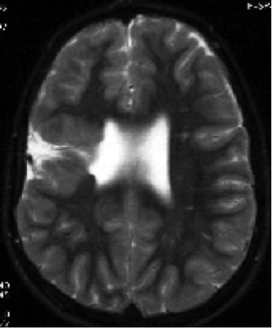
Imaging of a schizencephalic cleft
The image below shows a right sided open-lipped schizencephalic cleft. The walls of the cleft are lined by grey matter and are separated by CSF filled space.
Imaging characteristics of closed-lip schizencephaly:
- a cleft is seen extending from the ventricle to the cortical surface, composed of heterotopic grey matter
- a 'dimple' in the ventricle is seen, at the point where the cleft commences
- the cleft walls are in apposition, without CSF filled space separating them
- the closed-lip form is common in unilateral schizencephaly
Feedback
|
Home
|
Contact Us
|
Privacy
|
Terms & Conditions of Use
|
Log In For Videos

This website is owned by the International League Against Epilepsy. Text on this website, last updated June 30, 2024,
is available under a Creative Commons Attribution-ShareAlike 4.0 International License,
EXCEPTING all videos and images, which remain copyrighted by the International League Against Epilepsy.