- Overview
- Log In For Videos
- Give Feedback
- Seizure Classification
- Unknown Onset Seizure
- Neonatal Seizure
- Epilepsy Classification
- Generalized Epilepsy
- Focal Epilepsy
- Generalized and Focal Epilepsy
- Unknown Epilepsy
- Epilepsy Syndromes
- Epilepsy Etiologies
- Metabolic Etiologies
- Immune Etiologies
- Infectious Etiologies
- Unknown Etiologies
- Encephalopathy
- Epilepsy imitators
LISSENCEPHALY
Imaging
Imaging for optimized detection of lissencephaly:
Whilst lissencephaly may be seen on USS (antenatal and postnatal) and CT, MRI is the imaging of choice for assessing the detail and associated structural abnormalities. MRI should include thin slice volumetric T1-weighted images, axial and coronal T2-weighted and FLAIR images.
Imaging characteristics of lissencephaly type 1:
- reduced gyration (resulting in a shallow Sylvian fissure giving a 'figure of 8' appearance)
- cerebral cortical thickening, 12-20mm (instead of 3-4mm)
- subcortical band heterotopia is sometimes seen (anterior predominance in DCX pathogenic variants, posterior predominance in LIS1 pathogenic variants)
- other structural brain abnormalities may be seen such as enlarged ventricles and hypoplastic corpus callosum
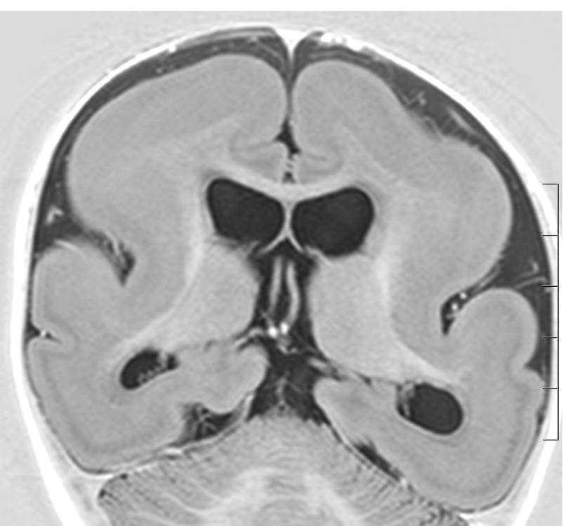
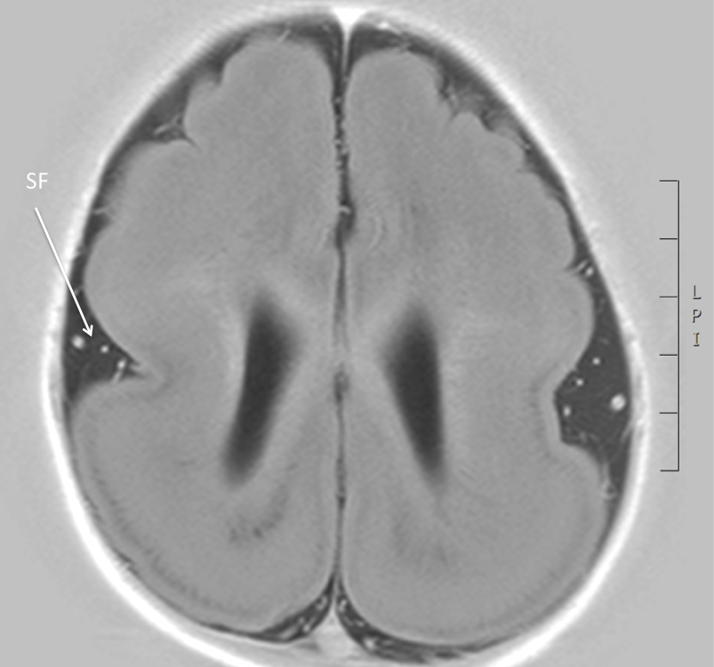
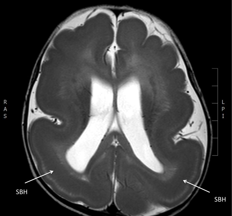
Imaging in lissencephaly type 1
The images below are all from the same patient. They show reduced gyration, a shallow Sylvian fissure (SF, arrow), thickening of the cortex and subcortical band heterotopia, appreciated over posterior regions (SBH, arrows).
Imaging characteristics of lissencephaly type 2 can include:
- lack of normal sulcation (resulting in a shallow Sylvian fissure giving a 'figure of 8' appearance)
- nodular cortical surface
- associated abnormalities, related to associated syndromes
Feedback
|
Home
|
Contact Us
|
Privacy
|
Terms & Conditions of Use
|
Log In For Videos

This website is owned by the International League Against Epilepsy. Text on this website, last updated June 30, 2024,
is available under a Creative Commons Attribution-ShareAlike 4.0 International License,
EXCEPTING all videos and images, which remain copyrighted by the International League Against Epilepsy.