- Overview
- Log In For Videos
- Give Feedback
- Seizure Classification
- Unknown Onset Seizure
- Neonatal Seizure
- Epilepsy Classification
- Generalized Epilepsy
- Focal Epilepsy
- Generalized and Focal Epilepsy
- Unknown Epilepsy
- Epilepsy Syndromes
- Epilepsy Etiologies
- Metabolic Etiologies
- Immune Etiologies
- Infectious Etiologies
- Unknown Etiologies
- Encephalopathy
- Epilepsy imitators
HYPOTHALAMIC HAMARTOMA
Imaging
Imaging for optimized detection of hypothalamic hamartoma:
Coronal T2 fast-spin echo sequences with thin slices and no inter-slice gap through the hypothalamus, to visualize the hypothalamic hamartoma and its attachment.
Imaging characteristics of hypothalamic hamartoma:
- a mass, of grey matter signal intensity, in the region of the hypothalamus
- may be pedunculated, attached to the mammillary region, and if large may distort or incorporate the mammillary bodies, with the columns of the fornix displaced antero-laterally and with variable extension below the third ventricle
- may be 'pedunculated', attached to the tuber cinereum, projecting into the suprasellar cistern
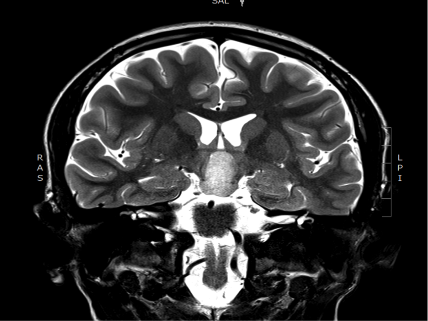
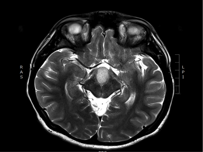
Imaging of a hypothalamic hamartoma
The images below show a hypothalamic hamartoma, seen on axial and coronal T2-weighted imaging, evident as a mass with signal that is slightly brighter than that of the cortex, attached to the mamillary bodies, and displacing the columns of the fornix laterally.
Feedback
|
Home
|
Contact Us
|
Privacy
|
Terms & Conditions of Use
|
Log In For Videos

This website is owned by the International League Against Epilepsy. Text on this website, last updated June 30, 2024,
is available under a Creative Commons Attribution-ShareAlike 4.0 International License,
EXCEPTING all videos and images, which remain copyrighted by the International League Against Epilepsy.