- Overview
- Log In For Videos
- Give Feedback
- Seizure Classification
- Unknown Onset Seizure
- Neonatal Seizure
- Epilepsy Classification
- Generalized Epilepsy
- Focal Epilepsy
- Generalized and Focal Epilepsy
- Unknown Epilepsy
- Epilepsy Syndromes
- Epilepsy Etiologies
- Metabolic Etiologies
- Immune Etiologies
- Infectious Etiologies
- Unknown Etiologies
- Encephalopathy
- Epilepsy imitators
HEMIMEGALENCEPHALY
Imaging
Imaging for optimized detection of hemimegalencephaly:
Although hemimegalencephaly may be identified on USS (fetal and postnatal) and CT, MRI is the imaging of choice for assessing the detail and associated structural abnormalities. MRI should include thin slice volumetric T1-weighted images, axial and coronal T2-weighted and FLAIR images.
Imaging characteristics of hemimegalencephaly:
- the cortex shows areas of thickening, with shallow sulci and enlarged/poorly formed gyri, it may also show areas of polymicrogyria, grey matter heterotopia or pachygyria
- the grey-white matter junction is blurred
- white matter volume is abnormal and demonstrates abnormal signal
- the appearance of the ventricle is abnormal, particularly the posterior horn of the lateral ventricle, as the occipital lobe may be displaced across the midline
- the cerebral enlargement may be maximal posteriorly, appearing as a posterior quadrantic dysplasia
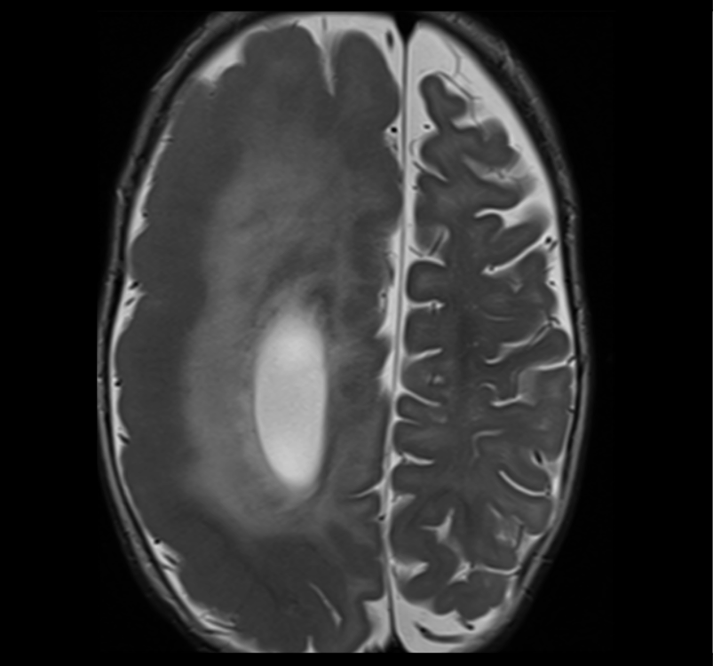
Imaging of hemimegalencephaly
The image below shows an enlarged right hemisphere, with thickened cortex with shallow sulcation, blurring of the grey-white matter junction and abnormal signal in the white matter.
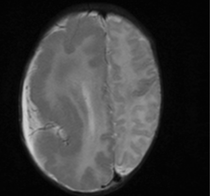
Imaging of hemimegalencephaly
The image below shows an enlarged right hemisphere, with thickened cortex, blurring of the grey-white matter junction and abnormal signal in the white matter.
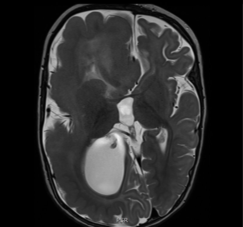
Imaging of hemimegalencephaly
The image below shows an enlarged right hemisphere, with thickened cortex, blurring of the grey-white matter junction and abnormal signal in the white matter. The right basal ganglia is abnormal in structure and orientation. Ventricular size and morphology is abnormal, and the occipital lobe extends across the midline.
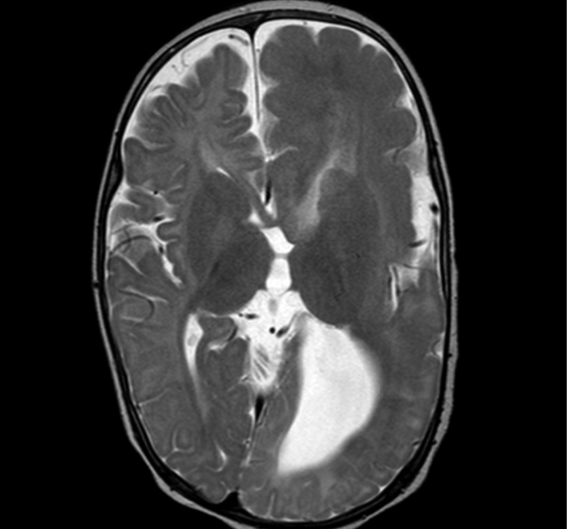
Imaging of hemimegalencephaly
The image below shows an enlarged left hemisphere, with thickened cortex, blurring of the grey-white matter junction and abnormal signal in the white matter. The left basal ganglia is abnormal in structure and orientation. Ventricular size and morphology is abnormal, and the occipital lobe extends across the midline.
This website is owned by the International League Against Epilepsy. Text on this website, last updated June 30, 2024,
is available under a Creative Commons Attribution-ShareAlike 4.0 International License,
EXCEPTING all videos and images, which remain copyrighted by the International League Against Epilepsy.