- Overview
- Log In For Videos
- Give Feedback
- Seizure Classification
- Unknown Onset Seizure
- Neonatal Seizure
- Epilepsy Classification
- Generalized Epilepsy
- Focal Epilepsy
- Generalized and Focal Epilepsy
- Unknown Epilepsy
- Epilepsy Syndromes
- Epilepsy Etiologies
- Metabolic Etiologies
- Immune Etiologies
- Infectious Etiologies
- Unknown Etiologies
- Encephalopathy
- Epilepsy imitators
FOCAL CORTICAL DYSPLASIA
Imaging
Imaging for optimized detection of focal cortical dysplasia:
MRI, with thin slice volumetric T1-weighted images, axial and coronal T2-weighted and FLAIR images.
Imaging characteristics of FCD type I:
- Atrophy - lobar or sub-lobar, with regional loss of subcortical white matter
- Increased signal in the white matter on T2-weighted/FLAIR imaging, and decreased signal on T1-weighted imaging
- May have mildly abnormal sulcal / gyral pattern
- FCD type Ia is usually found in the temporal lobes and may be associated with hippocampal atrophy, FCD type Ib is usually found outside of the temporal lobes
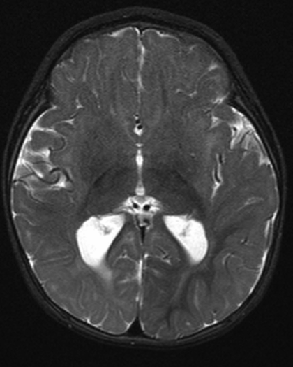
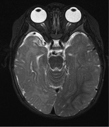
Imaging of FCD type I
Both images below are from the same patient and illustrate FCD type I, showing regional increase in signal in white matter (which appears brighter) on T2-weighted images, with regional atrophy (compare right temporal, parietal and occipital regions to the same regions on the left)
Imaging characteristics of FCD type II:
- Increased cortical thickness, with abnormal sulcal / gyral pattern
- Blurring of the grey-white matter junction
- Increased signal in the white matter on T2-weighted/FLAIR imaging, decreased signal on T1-weighted images
- A radially-oriented linear or conical transmantle stripe of T2/FLAIR hyperintensity tapering to the lateral ventricle
- FCD type II are most commonly found in the frontal lobes
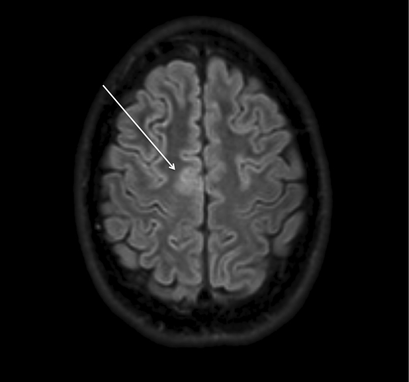
Imaging of FCD type II
There is a localized area of cortical thickening, with blurring of the grey-white matter junction and increased signal in the underlying white matter on FLAIR imaging, seen in the left frontal lobe
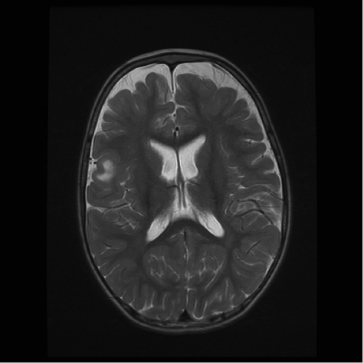
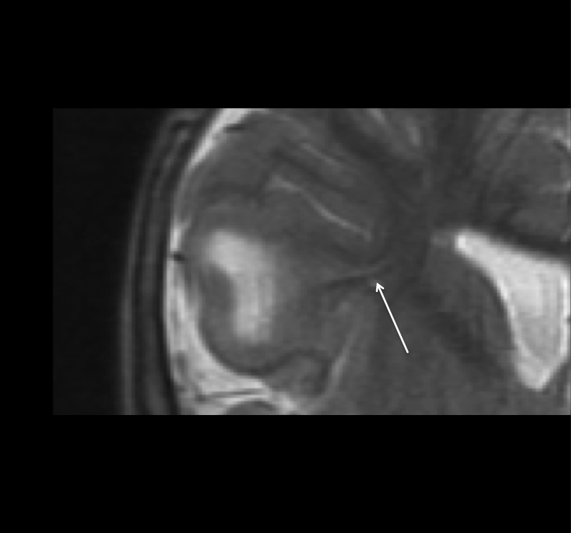
Imaging of FCD type II
There is a regional area of gyral expansion in the left inferior frontal lobe, with increased signal in the underlying white matter on T2-weighted imaging (with a central dark core) that is radially-orientated, tapering towards the lateral ventricle (with the 'tail' seen on the second coronal image, arrow). The imaging features FCD type II, can be identical to that of a cortical tuber.
CAUTION in the immature unmyelinated brain, increased T2/FLAIR signal is difficult to identify, as is grey-white matter junction blurring, imaging may need to be repeated after myelination is complete.
CAUTION some focal cortical dysplasias may be difficult to detect, but detection is important as epilepsy surgery can cure intractable seizures that arise from focal cortical dysplasias. Advanced imaging modalities, such as PET and SPECT scans, with expert review, may be required.
This website is owned by the International League Against Epilepsy. Text on this website, last updated June 30, 2024,
is available under a Creative Commons Attribution-ShareAlike 4.0 International License,
EXCEPTING all videos and images, which remain copyrighted by the International League Against Epilepsy.