- Overview
- Log In For Videos
- Give Feedback
- Seizure Classification
- Unknown Onset Seizure
- Neonatal Seizure
- Epilepsy Classification
- Generalized Epilepsy
- Focal Epilepsy
- Generalized and Focal Epilepsy
- Unknown Epilepsy
- Epilepsy Syndromes
- Epilepsy Etiologies
- Metabolic Etiologies
- Immune Etiologies
- Infectious Etiologies
- Unknown Etiologies
- Encephalopathy
- Epilepsy imitators
CEREBRAL ANGIOMA
Imaging
Imaging for optimized detection of cerebral angioma:
Gradient echo (GRE) or T2* sequences are able to delineate these lesions better than T1 or T2 weighted images, and GRE/T2* images are very important in detecting angiomas, that may be missed by conventional spin echo sequences. Susceptibility weighted imaging (SWI) may have sensitivity equal to that of GRE in detecting angiomas. SWI is also highly sensitive in detecting calcification as compared to T1 and T2 images. Unless large, or associated with hemorrhage, these abnormalities are not seen on CT. Cerebral angiomas are not seen on angiography, as they are 'slow flow' abnormalities.
Imaging characteristics of cerebral angioma:
- a mass with a characteristic "popcorn"" or "berry" appearance, with a rim of hemosiderin, which demonstrates prominent blooming on susceptibility weighted sequences
- T1 and T2 signal is varied internally due to degrading hemorrhage of varying age
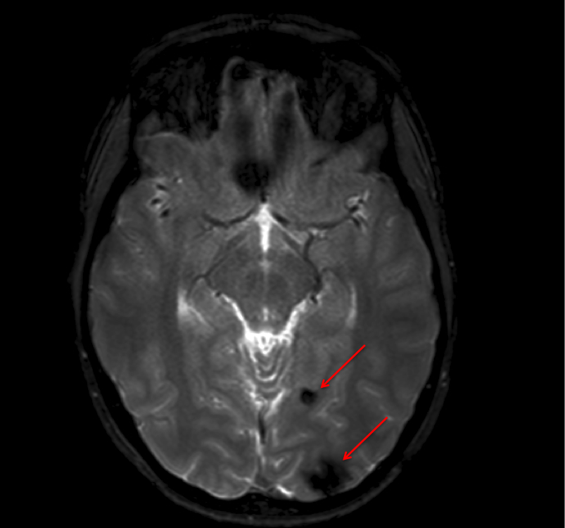
Imaging of cerebral angiomas
The image below, a T2* image, shows two punctate hypointense foci ('black dots') in the left occipital region (arrows), with others in the frontal lobes.
This website is owned by the International League Against Epilepsy. Text on this website, last updated June 30, 2024,
is available under a Creative Commons Attribution-ShareAlike 4.0 International License,
EXCEPTING all videos and images, which remain copyrighted by the International League Against Epilepsy.